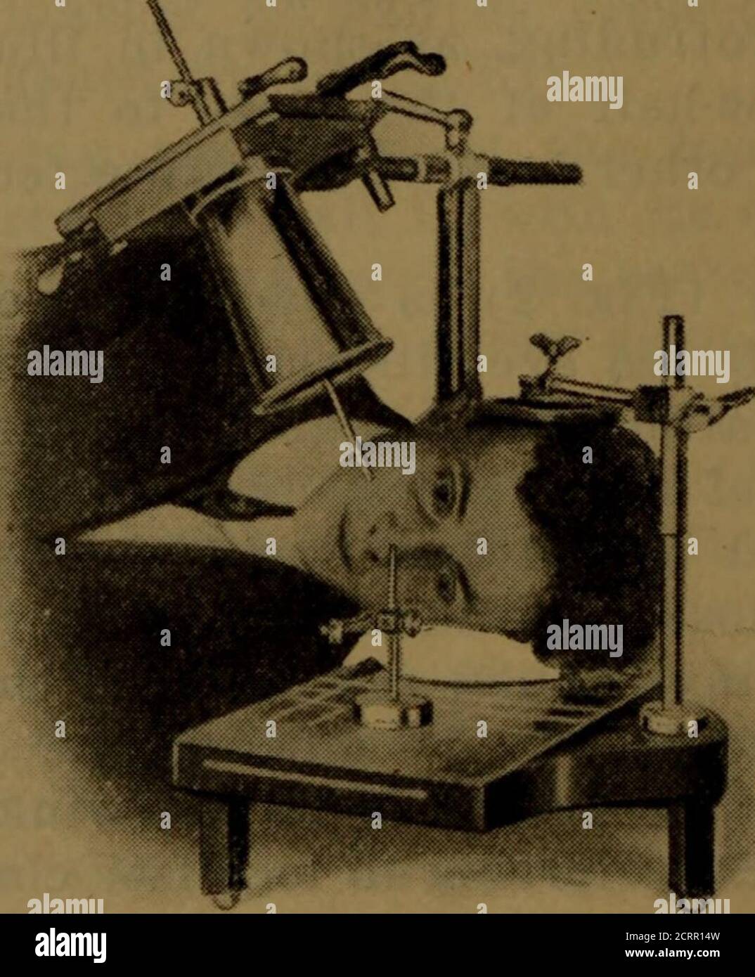
X-ray manual : U.S. Army . <*5 lit w Fig. 3. Position for first exposure.. Fig. 4. Position for second exposure.200 HEAD EXAMINATIONS 201 moved from the front plane of the
Download this stock image: . X-ray manual : U.S. Army . <*5 lit w Fig. 3. Position for first exposure.. Fig. 4. Position for second exposure.200 HEAD EXAMINATIONS 201 moved from the front plane of the cornea, and it shouldalso be borne in mind that the front of the cornea is 10millimeters in front of the shadow of the indicator-ball, asshown in your negatives. The tube is now centered overthe localizing ball and cone so that the shadows of thetwo will coincide (Fig. 3). Some object, such as a candle or a piece of whitepaper, that can be readily seen by the patient, should beplaced in alignment with the sights of the - 2CRR14W from Alamy's library of millions of high resolution stock photos, illustrations and vectors.
Radiographic Exposure Technique

Thermodynamics: An Engineering Approach - 5th Edition - Part II by 黑傑克 - Issuu

Radiological imaging of the Neonatal Chest by Online's Books - Issuu

2022 CPO Manual by thePHTA - Issuu

Operational guidelines and procedures for measuring the real size of the world economy part 1 by World Bank Publications - Issuu

unified facilities criteria (ufc)

Catalogo Completo by Hensistemas Hensistemas - Issuu

Al mefty meningiomas by Neurocirurgiao bh - Dr Eric Grossi - Issuu

Principles of Naval Weapons Systems by Francisco Azevedo - Issuu

United States Army X-Ray Manual: United States. Surgeon-General's Office: 9781141999200: : Books

PDF) X-ray solution scattering (SAXS) combined with crystallography and computation: defining accurate macromolecular structures, conformations and assemblies in solution

Essentials.of.Dental.Radiography.and.Radiology by Alejandro Padilla - Issuu

Miscellaneous - Clark's Positioning In Radiography - by A. S. Whitley

IJSPT Volume 17 Number 2 by IJSPT - Issuu